
What is Mohs Skin Cancer Surgery?
Mohs Micrographic Surgery is a specialised, precision surgical technique used for the treatment of skin cancer. It combines surgical excision with complete microscopic margin analysis.
International skin cancer guidelines recognise Mohs surgery as the first-line treatment and gold standard for skin cancers at high risk of recurrence, as well as those located in cosmetically sensitive or functionally important areas. It offers the highest cure rates (up to 99%), is cost-effective, and is associated with the best cosmetic outcomes.
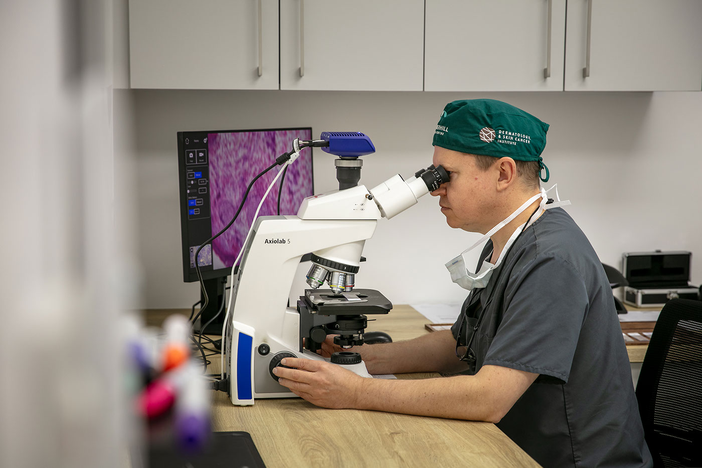
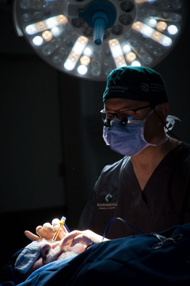
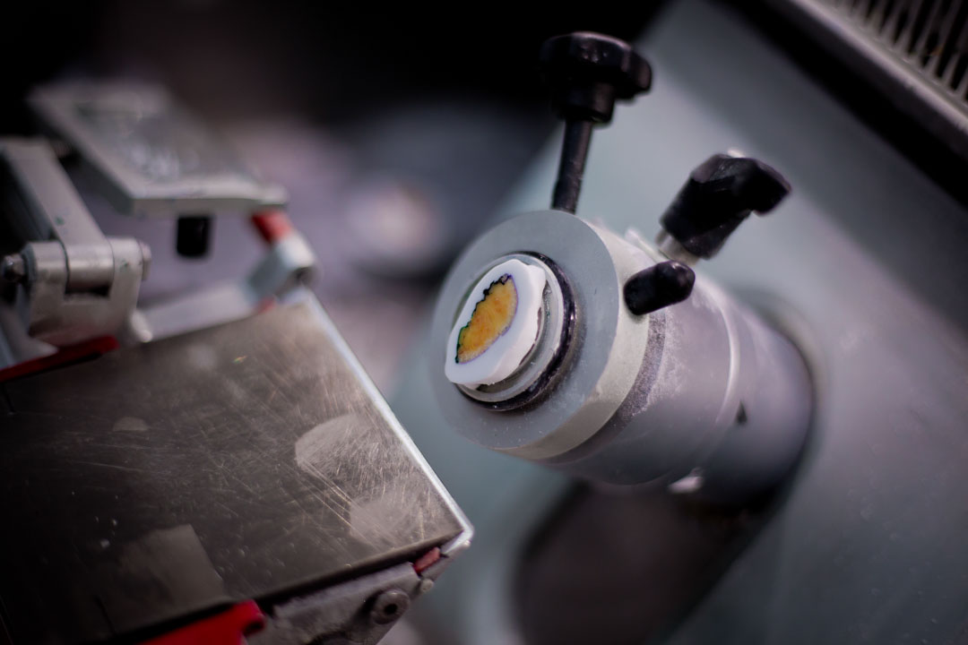
Mohs skin cancer surgery is usually performed under local anaesthetic in a dedicated day theatre, such as Summerhill Surgical Centre in Somerset West, near Cape Town. During the procedure, the tumour is removed together with a thin margin of surrounding healthy tissue. The excised tissue is processed immediately on site in a Mohs histological laboratory, while the patient returns to the ward.
The Mohs-specific method of tissue processing produces slides that show 100% of the peripheral and deep margins around and beneath the tumour. The Mohs surgeon then examines these slides to determine whether any cancer cells remain. (See Mohs surgeons and members of the South African Mohs Surgery Society.)
If residual tumour is identified, the patient returns to the theatre for a further excision – only in the precise area where tumour remains, preserving as much healthy tissue as possible. This results in the smallest possible surgical defect.
Once the Mohs surgeon confirms that the cancer has been completely removed, the wound is repaired either by a specialist reconstructive surgeon or by the Mohs surgeon.
The result of Mohs surgery is the highest cure rate, with maximum tissue preservation, and often a simpler reconstruction.
Dr Johann de Wet offers Mohs surgery to suitable candidates in Stellenbosch, Somerset West, and the greater Cape Town area. He is a fellowship-trained Mohs surgeon and forms part of the Skinmatters Mohs and Reconstructive Unit.
What to expect on the day of Mohs surgery:
- During your initial consultation the Mohs surgery will be discussed in detail.
- Plan to spend the whole day with us.
- The morning will be dedicated to the excision of the tumour and the analysis of the tissue.
- The excision will be done under local anaesthetic.
- The reconstruction of the defect will only take place in the afternoon once it has been confirmed that the skin cancer is completely removed.
- This is typically done under conscious sedation.
- Detailed instructions will be given to you by the reconstructive team regarding wound care and follow-up.
How does Mohs skin cancer surgery differ from traditional skin cancer surgery?
In conventional skin cancer surgery, the tumour is removed together with a predetermined margin of normal skin — often more than 5 mm for high-risk basal cell carcinomas (BCC) and over 10 mm for squamous cell carcinomas (SCC). Although this strategy aims to clear the cancer, it can create unnecessarily large wounds and sometimes requires more complex reconstruction. A key limitation is that the surgeon has no immediate confirmation that all cancer cells have been removed, which means that despite removing a generous amount of healthy tissue, residual cancer may still be left behind.
With standard excision, the specimen is usually evaluated using vertical or “bread-loaf” sectioning, where only a very small fraction of the true margin (approximately 1%) is examined under the microscope. By comparison, Mohs Micrographic Surgery allows for the complete assessment of 100% of the peripheral and deep margins, providing a far more accurate evaluation.
Because traditional histology examines only a limited sample, there is a higher chance that remaining tumour cells go undetected, which can lead to recurrence of the skin cancer over time.
The Mohs Surgery Process:
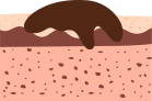
Step 1
Skin cancers often extend beyond their visible borders, and these extensions (roots) can cause the tumour to recur if not completely removed.
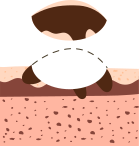
Step 2
The Mohs technique begins by removing the visible tumor with thin layer of healthy tissue.
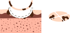
Step 3
This tissue is mapped and examined under a microscope by the surgeon to check for any cancerous cells.
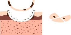
Step 4
If cancerous tissue remains, additional thin layers are removed, precisely focusing on the areas where the cancer persists, until the tumor is entirely eliminated.
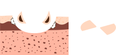
Step 5
Mohs surgery is unique in that it ensures the entire tumor is removed while preserving the maximum amount of healthy tissue. This minimizes scarring and reduces the risk of cancer recurrence.